








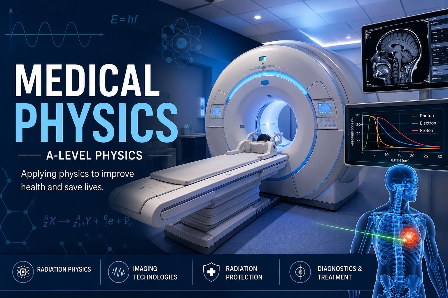
Medical Physics is one of the most fascinating and rewarding topics in A-Level Physics because it demonstrates how the principles of physics directly improve human health and save lives. Every day, hospitals around the world rely on technologies developed through physics to diagnose illnesses, monitor patients, and treat life-threatening diseases.
Whether a doctor is using an X-ray to identify a broken bone, an MRI scanner to examine the brain, an ultrasound scan to monitor the development of a baby, or a PET scanner to detect cancer, physics is working behind the scenes. Medical physicists collaborate with doctors, radiographers, engineers, and researchers to ensure that these technologies provide accurate diagnoses while keeping patients safe from unnecessary radiation exposure.
For A-Level Physics students, Medical Physics is much more than memorising different scanning techniques. It is about understanding how electromagnetic waves, sound waves, nuclear radiation, magnetism, electricity, and modern computing combine to create powerful medical technologies that have transformed healthcare.
In this comprehensive guide, we will explore the major medical imaging techniques studied in A-Level Physics, explain the physics behind each method, compare their advantages and disadvantages, discuss radiation safety, and provide examination tips to help you achieve top grades.
Medical Physics is the application of physics principles to medicine. It combines several areas of physics, including:
Electromagnetic radiation
Nuclear physics
Wave behaviour
Magnetism
Electronics
Computer science
Signal processing
Medical physicists work to improve the diagnosis and treatment of disease by developing safer and more accurate imaging technologies. They also calculate radiation doses for cancer treatments and ensure that medical equipment operates correctly and safely.
Without physics, modern medicine would be dramatically less effective.
Many diseases cannot be diagnosed simply by looking at a patient. Doctors need to see inside the body without performing surgery whenever possible.
Medical imaging allows doctors to:
Detect broken bones
Diagnose cancer
Identify internal bleeding
Examine the brain
Investigate heart disease
Monitor unborn babies
Locate infections
Plan surgical procedures
Different imaging methods are suited to different parts of the body because they rely on different physical principles.
One of the most important concepts in Medical Physics is understanding the difference between ionising and non-ionising radiation.
Ionising radiation carries enough energy to remove electrons from atoms, producing ions. Because ionisation can damage DNA, excessive exposure increases the risk of cancer.
Examples include:
X-rays
Gamma rays
Alpha particles
Beta particles
Medical professionals carefully control patient exposure to ionising radiation.
Non-ionising radiation does not have sufficient energy to remove electrons from atoms.
Examples include:
Radio waves
Microwaves
Infrared radiation
Visible light
Ultrasound
MRI radio-frequency waves
These techniques are generally considered much safer for repeated use.
X-rays are high-energy electromagnetic waves with wavelengths much shorter than visible light. Their high frequency gives them enough energy to penetrate soft tissues while being absorbed more strongly by denser materials such as bone.
This difference in absorption creates an image.
Inside an X-ray tube:
A heated filament releases electrons.
A high voltage accelerates the electrons.
Electrons strike a tungsten target.
Their kinetic energy is converted into X-rays.
Only a small percentage of the energy becomes X-rays. Most is converted into heat.
Bones contain calcium, which has a relatively high atomic number and density. As a result, bones absorb more X-rays than surrounding soft tissue.
Bone → absorbs many X-rays → appears white
Muscle → absorbs fewer X-rays → appears grey
Air → absorbs almost none → appears black
Fast
Relatively inexpensive
Excellent for detecting fractures
Widely available
Uses ionising radiation
Poor soft tissue contrast
Provides only a two-dimensional image
A CT scanner is an advanced form of X-ray imaging.
Instead of taking a single picture, the X-ray source rotates around the patient while detectors measure the transmitted radiation from many different angles.
A computer reconstructs these measurements into detailed cross-sectional images.
Produces detailed three-dimensional images
Excellent for internal organs
Detects bleeding and tumours
Useful in emergency medicine
Higher radiation dose than ordinary X-rays
More expensive
Longer scanning time
Unlike X-rays, ultrasound uses high-frequency sound waves rather than electromagnetic radiation.
The transducer contains piezoelectric crystals.
These crystals convert electrical energy into sound waves and convert returning echoes back into electrical signals.
The computer analyses the time taken for echoes to return to determine the depth of different tissues.
Ultrasound is ideal for:
Pregnancy scans
Heart examinations
Liver imaging
Kidney investigations
Blood flow measurements
No ionising radiation
Safe for unborn babies
Real-time imaging
Portable
Relatively inexpensive
Cannot pass through bone
Air interferes with imaging
Lower resolution than CT or MRI
MRI uses one of the most sophisticated technologies in modern medicine.
Unlike CT scanners, MRI does not use ionising radiation.
Instead, it combines:
Extremely strong magnetic fields
Radio-frequency pulses
Advanced computer processing
Hydrogen nuclei (protons) inside the body behave like tiny magnets.
When placed inside a strong magnetic field, they align with that field.
Radio waves disturb this alignment.
As the protons return to their original state, they emit radio-frequency signals.
These signals are detected and used to construct detailed images.
Excellent soft tissue contrast
No ionising radiation
Ideal for brain imaging
Excellent for spinal injuries
High image quality
Expensive
Slow scanning
Cannot be used with some metal implants
Claustrophobic for some patients
PET scanning studies how organs function rather than simply showing their structure.
A radioactive tracer is injected into the patient.
The tracer emits positrons.
Each positron quickly annihilates with an electron, producing two gamma photons travelling in opposite directions.
Detectors surrounding the patient detect both photons simultaneously.
Computers reconstruct the location where annihilation occurred.
PET is especially useful for:
Cancer diagnosis
Alzheimer's disease
Brain activity
Heart function
Gamma cameras detect gamma rays emitted from radioactive tracers inside the body.
They are widely used in nuclear medicine to examine:
Thyroid gland
Kidneys
Bones
Heart
Liver
Unlike X-rays, the radiation originates inside the patient rather than outside.
Radiotherapy is one of the most important treatments for cancer.
High-energy radiation damages the DNA of cancer cells.
Because cancer cells divide rapidly, they are less able to repair this damage than healthy cells.
Modern radiotherapy carefully shapes the radiation beam so that the tumour receives the highest dose while healthy tissues receive as little radiation as possible.
Types include:
External beam radiotherapy
Brachytherapy
Proton therapy
Medical physicists measure radiation dose using several quantities.
Measured in:
Gray (Gy)
1 Gy = 1 joule of energy absorbed per kilogram of tissue.
Accounts for the biological effects of different radiation types.
Measured in:
Sievert (Sv)
Radiation protection follows the ALARA principle:
As Low As Reasonably Achievable
Three important methods reduce radiation exposure:
Reduce exposure time.
Increase distance from the radiation source.
Use lead or concrete barriers.
| Technique | Radiation | Best For | Main Advantage |
|---|---|---|---|
| X-ray | Ionising | Bones | Fast and inexpensive |
| CT | Ionising | Internal organs | Detailed 3D images |
| MRI | Non-ionising | Soft tissue | Excellent contrast |
| Ultrasound | Non-ionising | Pregnancy | Safe and portable |
| PET | Ionising | Metabolism | Detects disease activity |
Students are often asked to:
Compare MRI and CT scanners.
Explain why ultrasound is safe during pregnancy.
Describe how CT scanners produce three-dimensional images.
Explain why lead is used for shielding.
Discuss the risks and benefits of ionising radiation.
Many students lose marks by:
Confusing MRI with CT scanning.
Forgetting that ultrasound uses sound rather than electromagnetic waves.
Assuming all radiation is dangerous.
Mixing absorbed dose (Gray) with equivalent dose (Sievert).
Forgetting that PET scans show function rather than structure.
To achieve high marks:
Learn the advantages and disadvantages of every imaging technique.
Understand how each scanner works rather than memorising facts.
Practise comparison questions.
Know which techniques use ionising radiation.
Revise radiation safety principles.
Learn common medical applications of each method.
Medical Physics continues to evolve rapidly. Artificial intelligence is now helping doctors interpret medical images more accurately, while proton therapy is improving cancer treatment by delivering radiation with greater precision. Researchers are also developing portable MRI systems, advanced PET scanners, and hybrid imaging technologies that combine multiple scanning methods to provide even more detailed information.
Medical Physics demonstrates the incredible impact that physics has on healthcare. From diagnosing broken bones with X-rays to detecting cancer with PET scanners and treating tumours using radiotherapy, the application of physics has transformed modern medicine.
For A-Level Physics students, understanding the principles behind medical imaging is essential not only for examination success but also for appreciating how scientific knowledge improves millions of lives every year. By mastering the advantages, limitations, and physical principles of each imaging technique, you'll be well prepared for exam questions and gain a deeper understanding of one of the most rewarding applications of physics.
Join our GCSE and A-Level Physics tutoring programmes and discover how expert guidance can help you achieve outstanding examination results.
Book Your Free Assessment





.png)

.png)
.png)
.png)




.png)
.png)

.png)


.png)





.png)


.png)
1.png)






.jpeg)





See our Trustpilot Excellent Reviews